Home
Understanding the Electrocardiogram

Main navigation

  • Home
  • Understanding ECGs
    • Introduction
User account menu
  • Log in

Breadcrumb

  1. Home
  2. Understanding the Electrocardiogram In Health & Disease
  3. 4.0.0 Ventricular Hypertrophy
  4. 4.3.0 Biventricular hypertrophy (frame63)

4.3.4(frame 67)

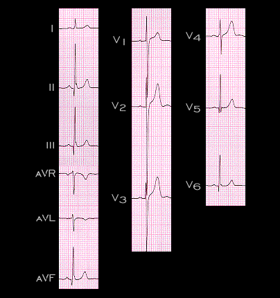
The ECG shown here is from a 19 year old male with a loud systolic murmur heard best in the 4th left intercostal space. The electrocardiogram, with the deep S waves in leads V2 and V3, is still within normal limits for a 19 year old male. The loud murmur reflected a high velocity of flow across a small ventricular septal defect. There was no ventricular hypertrophy and the pulmonary artery pressure was normal. This constellation of findings, namely a small VSD with a loud murmur and a normal ECG, is referred to as "Maladie de Roger" after the 19th century French pediatrician who first described it.

Book traversal links for 4.3.4(frame 67)

  • 4.3.3 (frame 66)
  • Up
  • 5.0.0 ELECTROLYTE ABNORMALITIES, DRUG EFFECTS AND THE LONG QT SYNDROMES

Site is under construction

Book navigation

  • Introduction to First Edition
  • 1.0.0 Generation of the ECG
  • 2.0.0 The Normal Electrocardiogram
  • 3.0.0 Inter and intra-ventricular Conduction Disturbances
  • 4.0.0 Ventricular Hypertrophy
    • 4.1.0 Left Ventricular Hypertrophy (frame 1)
    • 4.2.0 Right Ventricular Hypertrophy (frame 21)
    • 4.3.0 Biventricular hypertrophy (frame63)
      • 4.3.1 (frame 64)
      • 4.3.2 (frame 65
      • 4.3.3 (frame 66)
      • 4.3.4(frame 67)
  • 5.0.0 ELECTROLYTE ABNORMALITIES, DRUG EFFECTS AND THE LONG QT SYNDROMES
  • 6.0.0 Ischemia and Infarction - Introduction (frame i and ii)
  • 7.0.0 Tachycardias- Introduction
  • 8.0.0 The Bradycardias frame i-introduction
  • 9.0.0 The ECG of Heart Murmurs-introduction
  • 10.0.0 The Electrocardiogram in the Emergency Department-Introduction
Powered by Drupal