
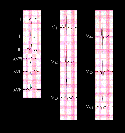
Patients with ventricular septal defects may also develop biventricular hypertrophy. The left ventricle is hypertrophied due to the increase in blood volume while the right is hypertrophied due to an increase in pulmonary artery pressure. The ECG shown here is from a 33 year old male with a large ventricular septal defect and moderately severe pulmonary hypertension. The right axis deviation (+115 degrees) and the tall R waves in leads V1 and V2 reflect the right ventricular hypertrophy while the deep S waves in leads V2 and V3 and the well preserved R waves in leads V5 and V6 reflect the left ventricular hypertrophy. This ECG pattern, with the large equiphasic QRS complexes in the mid precordial leads, is characteristic of the biventricular hypertrophy which accompanies ventricular septal defects and is referred to as the Katz-Wachtel pattern.