Home
Understanding the Electrocardiogram

Main navigation

  • Home
  • Understanding ECGs
    • Introduction
User account menu
  • Log in

Breadcrumb

  1. Home
  2. Understanding the Electrocardiogram In Health & Disease
  3. 4.0.0 Ventricular Hypertrophy
  4. 4.3.0 Biventricular hypertrophy (frame63)

4.3.3 (frame 66)

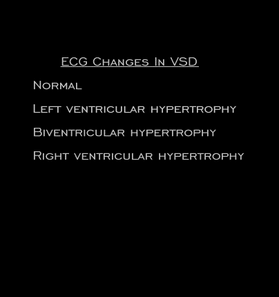
The electrocardiographic changes in patients with ventricular septal defects will depend on the magnitude of the shunt, and the presence (or absence), of pulmonary hypertension. For that reason, the electrocardiogram may be normal, or may indicate left venricular, right ventricular or biventricular hypertrophy.

Book traversal links for 4.3.3 (frame 66)

  • 4.3.2 (frame 65
  • Up
  • 4.3.4(frame 67)

Site is under construction

Book navigation

  • Introduction to First Edition
  • 1.0.0 Generation of the ECG
  • 2.0.0 The Normal Electrocardiogram
  • 3.0.0 Inter and intra-ventricular Conduction Disturbances
  • 4.0.0 Ventricular Hypertrophy
    • 4.1.0 Left Ventricular Hypertrophy (frame 1)
    • 4.2.0 Right Ventricular Hypertrophy (frame 21)
    • 4.3.0 Biventricular hypertrophy (frame63)
      • 4.3.1 (frame 64)
      • 4.3.2 (frame 65
      • 4.3.3 (frame 66)
      • 4.3.4(frame 67)
  • 5.0.0 ELECTROLYTE ABNORMALITIES, DRUG EFFECTS AND THE LONG QT SYNDROMES
  • 6.0.0 Ischemia and Infarction - Introduction (frame i and ii)
  • 7.0.0 Tachycardias- Introduction
  • 8.0.0 The Bradycardias frame i-introduction
  • 9.0.0 The ECG of Heart Murmurs-introduction
  • 10.0.0 The Electrocardiogram in the Emergency Department-Introduction
Powered by Drupal