
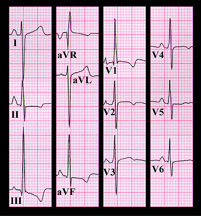
Her ECG is shown again here. The rhythm is sinus and the PR, QRS and QT intervals are normal. The frontal plane QRS spatial vector is directed inferiorly and to the right with an axis of +120 degrees. The tall P waves in leads II, II and aVF meet the criteria for "P pulmonale" and suggest right atrial enlargement. The qR complex with the tall R wave in lead V1, and the R and S waves of almost equal ampltude in V6 suggest a pressure overloaded right ventricle with marked right ventricular hypertrophy. The tracing is quite similar to that obtained from the patient with severe pulmonic stenosis (pages 9.1.10 and 9.1.11). However, the murmur in this patient is not the murmur one would expect with pulmonic stenosis or with an intra-cardiac shunt. These would be systolic murmurs. Rather, the diastolic murmur in this patient, in association with this electrocardiogram, suggests insufficiency of the pulmonic valve, the result of severe pulmonary hypertension caused by the sarcoidosis.