Home
Understanding the Electrocardiogram

Main navigation

  • Home
  • Understanding ECGs
    • Introduction
User account menu
  • Log in

Breadcrumb

  1. Home
  2. Understanding the Electrocardiogram In Health & Disease
  3. 7.0.0 Tachycardias- Introduction
  4. 7.1.0 Mechanisms of arrhythmias-frame 1
  5. 7.1.30 (frame 30)

7.1.31 (31)

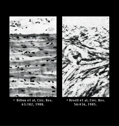
In the chronically diseased ventricle, regions of myocardial fibrosis and cells with altered electrophysiologic properties provide the substrate for re-entry. Shown here are photomicrographs of canine epicardial cells. The panel on the left was obtain a week after acutely ligating a coronary artery to cause a myocardial infarction and shows parallel myocardial fibers, slightly separated by inflammatory cells and edema. The panel on the right, from an earlier work, shows a similar section two months after the infarction. There are now areas of fibrosis and disarranged but viable myocardial cells capable of resulting in marked electrical inhomogeniety, altered conduction pathways, regions of slowed conduction and unidirectional block.


Book traversal links for 7.1.31 (31)

  • 7.1.30 (frame 30)
  • Up
  • 7.1.32 (32)

Site is under construction

Book navigation

  • Introduction to First Edition
  • 1.0.0 Generation of the ECG
  • 2.0.0 The Normal Electrocardiogram
  • 3.0.0 Inter and intra-ventricular Conduction Disturbances
  • 4.0.0 Ventricular Hypertrophy
  • 5.0.0 ELECTROLYTE ABNORMALITIES, DRUG EFFECTS AND THE LONG QT SYNDROMES
  • 6.0.0 Ischemia and Infarction - Introduction (frame i and ii)
  • 7.0.0 Tachycardias- Introduction
    • 7.1.0 Mechanisms of arrhythmias-frame 1
      • 7.1.1 (2)
      • 7.1.2 (3)
      • 7.1.3 (4)
      • 7.1.4 (5)
      • 7.1.5 (6)
      • 7.1.6 (7)
      • 7.1.7 (8)
      • 7.1.8 (9)
      • 7.1.9 (10)
      • 7.1.10 (11)
      • 7.1.20 (frame 20)
      • 7.1.30 (frame 30)
        • 7.1.31 (31)
        • 7.1.32 (32)
        • 7.1.33 (33)
        • 7.1.34 (34)
        • 7.1.35 (35)
        • 7.1.36 (36)
        • 7.1.37 (37)
    • 7.2.0 Narrow Complex Rhythms (frame 38)
    • 7.3.0 Atrial Flutter and Fibrillation (frame 93)
    • 7.4.0 Wide Complex Beats and Rhythms (frame 122)
  • 8.0.0 The Bradycardias frame i-introduction
  • 9.0.0 The ECG of Heart Murmurs-introduction
  • 10.0.0 The Electrocardiogram in the Emergency Department-Introduction
Powered by Drupal